


Subscribe To Get Information, Latest News And Other Interesting Events!


Important Links
Get In Touch
Ariston Publication
3167 Riverview Place
Columbus, OH 43202
United States of America
Copyright © 2026 Ariston Publications. All Rights Reserved.
Loading Your Library
Preparing your reading experience...
Loading content...
Ariston Publication
3167 Riverview Place
Columbus, OH 43202
United States of America

Ftema W. Aldbea, Salma Alsanousi Mannaa, Salem M. Aldosari, Nourah Mohammed Salih, Ali Adam Ali, Ali Basheer Yousuf, A. Sharma, Pramod K. Singh
Ftema W. Aldbea1,*
Salma Alsanousi Mannaa1
Salem M. Aldosari2
Nourah Mohammed Salih1
Ali Adam Ali3
Ali Basheer Yousuf3
A. Sharma4
Pramod K. Singh5
1 Physics department, Faculty of Science, Sebha University, Libya.
2 Material Science Research Institute, King Abdulaziz City for Science and Technology (KACST), Riyadh 11442, Saudi Arabia.
3 Department of Chemical Engineering, Faculty of Engineering, Sebha University, Libya.
4 Metal Extraction and Recycling Division (MER), CSIR-National Metallurgical Laboratory, Jamshedpur-831007, India.
5 Centre of Excellence in Solar Cell and Renewable Energy, Department of Physics and Environmental Sciences, Sharda School of Engineering and Science (SSES), Sharda University, Greater Noida, Uttar Pradesh, 201310, India.
* Author to whom correspondence should be addressed:
fte.aldbea@sebhau.edu.ly (Ftema W. Aldbea)
Pure and Mg-doped zinc sulfide nanoparticles (MgxZn1−xS, where x = 0.0 and 0.004) were prepared using a sol–gel method, followed by calcination in air at 200 °C for 2 h. The structural, morphological, elemental, and optical characteristics of the resulting materials were examined using XRD, FE-SEM combined with EDS, and UV–visible spectroscopy. XRD analysis showed that both samples have a cubic ZnS structure; however, in the doped material, minor MgS and MgO impurity phases appeared because the calcination temperature was not high enough to achieve complete crystallization. After Mg doping, the diffraction peaks moved to higher 2θ angles, in line with the lattice parameter, confirming that Mg atoms were incorporated into the ZnS nanoparticles. Furthermore, doping led to an increase in the average crystallite size, reaching 2.72 nm, as well as in the microstrain, which arose from the impurity phases and lattice distortion. An increase in the lattice parameter (from 5.38 to 5.39 Å) and in the unit cell volume (from 155.912 to 156.320 Å3) was also noted. FE-SEM analysis revealed that the pure ZnS powder was composed of agglomerated grains, while the doped sample exhibited stronger agglomeration and porosity, with visible voids. EDS analysis verified the presence of Zn and S in the pure ZnS and of Zn, S, and Mg in the doped sample. The reflectance edge was observed to shift from roughly 367 nm for pure ZnS to about 206 nm after the addition of Mg. In addition, the absorption edge shifted toward the visible region, and the optical energy gap declined from 3.6 to 1.8 eV. This narrowing of the band gap is a consequence of lattice expansion together with the presence of defects and impurity phases. Based on the calculated optical energy gap, it can be proposed that Mg-doped zinc sulfide nanoparticles may hold potential for applications in photolytic processes.
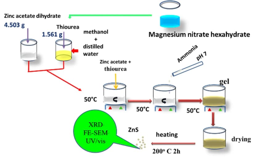
This work is significant because it demonstrates how magnesium doping modifies the structural, morphological, and optical properties of ZnS nanoparticles synthesized through a simple sol–gel process. Mg incorporation influences lattice parameters, crystallite size, microstrain, and porosity while substantially narrowing the optical band gap from 3.6 to 1.8 eV. This optical modification suggests potential for visible-light-driven photocatalytic applications. The findings provide valuable insights into composition–structure–property relationships and highlight the importance of optimizing calcination conditions to minimize impurity phases and improve material performance.
This study investigates pure and Mg-doped ZnS nanoparticles prepared by sol–gel synthesis and calcination at 200 °C. XRD confirmed cubic ZnS, with Mg doping producing minor MgS and MgO phases, increased crystallite size, and slight lattice expansion. FE-SEM revealed enhanced agglomeration and porosity, while EDS verified magnesium incorporation. Optical measurements showed a reflectance-edge shift from approximately 367 to 206 nm and a band-gap reduction from 3.6 to 1.8 eV, indicating potential for photocatalytic applications.
Article type: Research Article
Submitted: 21 June 2026
Revised: 1 July 2026
Accepted: 19 July 2026
First published: 15 August 2026
DOI: https://doi.org/10.69626/mse.2026.0147
Volume: 3
Issue: 03
Pages: 147-154
Citation: MatSci Express 3(3), 147-154 (2026)
Aldbea F. W., Mannaa S. A., Aldosari S. M., Salih N. M., Ali A. A., Yousuf A. B., Sharma A., Singh P. K. 2026. Structural and Optical Properties of MgxZn1-xS (x = 0.0 and 0.004) Nanoparticles. MatSci Express 3(3), pp. 147-154; https://doi.org/10.69626/mse.2026.0147
Number of downloads: 6
Number of views: 55